

Exams
Ultra low-dose chest scanner
Ultra low-dose chest CT scan: An advanced imaging technique that uses a radiation dose 20 times lower than that of a conventional CT scan, while maintaining optimal image quality.
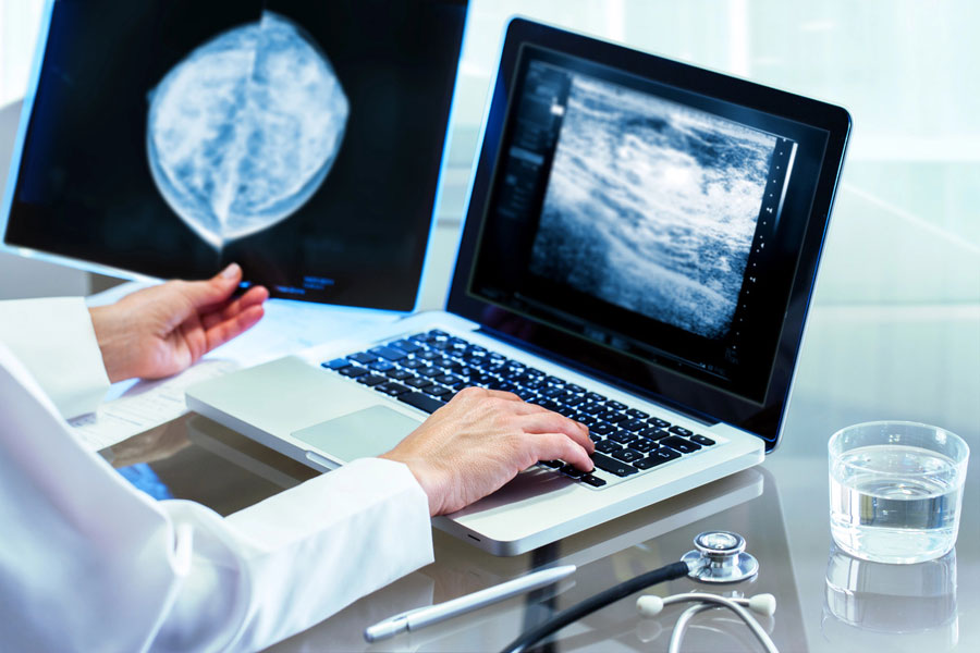
4D spectral mammography
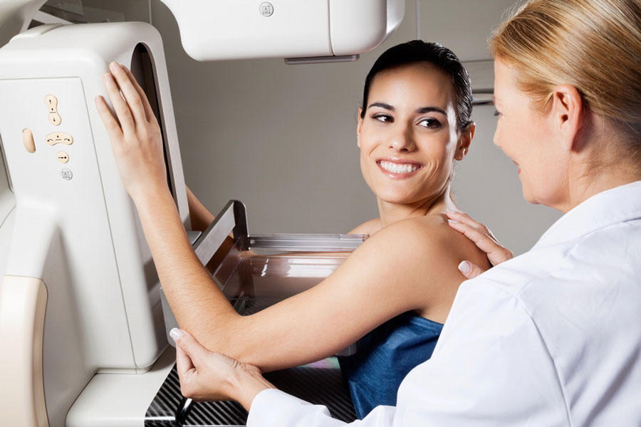
Innovative imaging technique that combines four-dimensional images with spectral analysis to improve the detection and characterization of breast abnormalities. This method allows you to better visualize the internal structures of the breast and differentiate between tissue types with more precision.
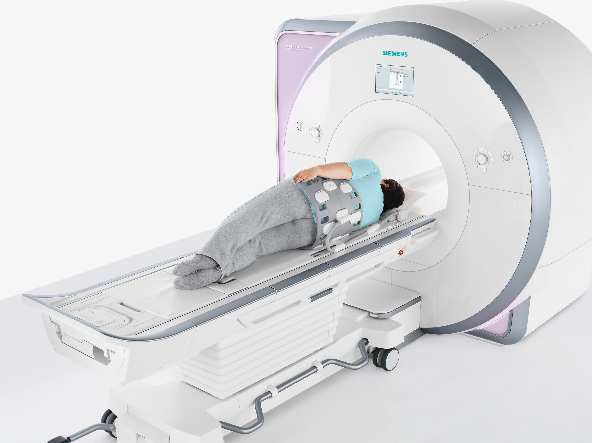
Prostate MRI

Prostate MRI is an advanced imaging examination that uses a powerful magnetic field and radio waves to obtain detailed images of the prostate and surrounding tissues. This examination is essential for the early detection of prostate cancer, the assessment of tumor extent, and post-treatment monitoring.
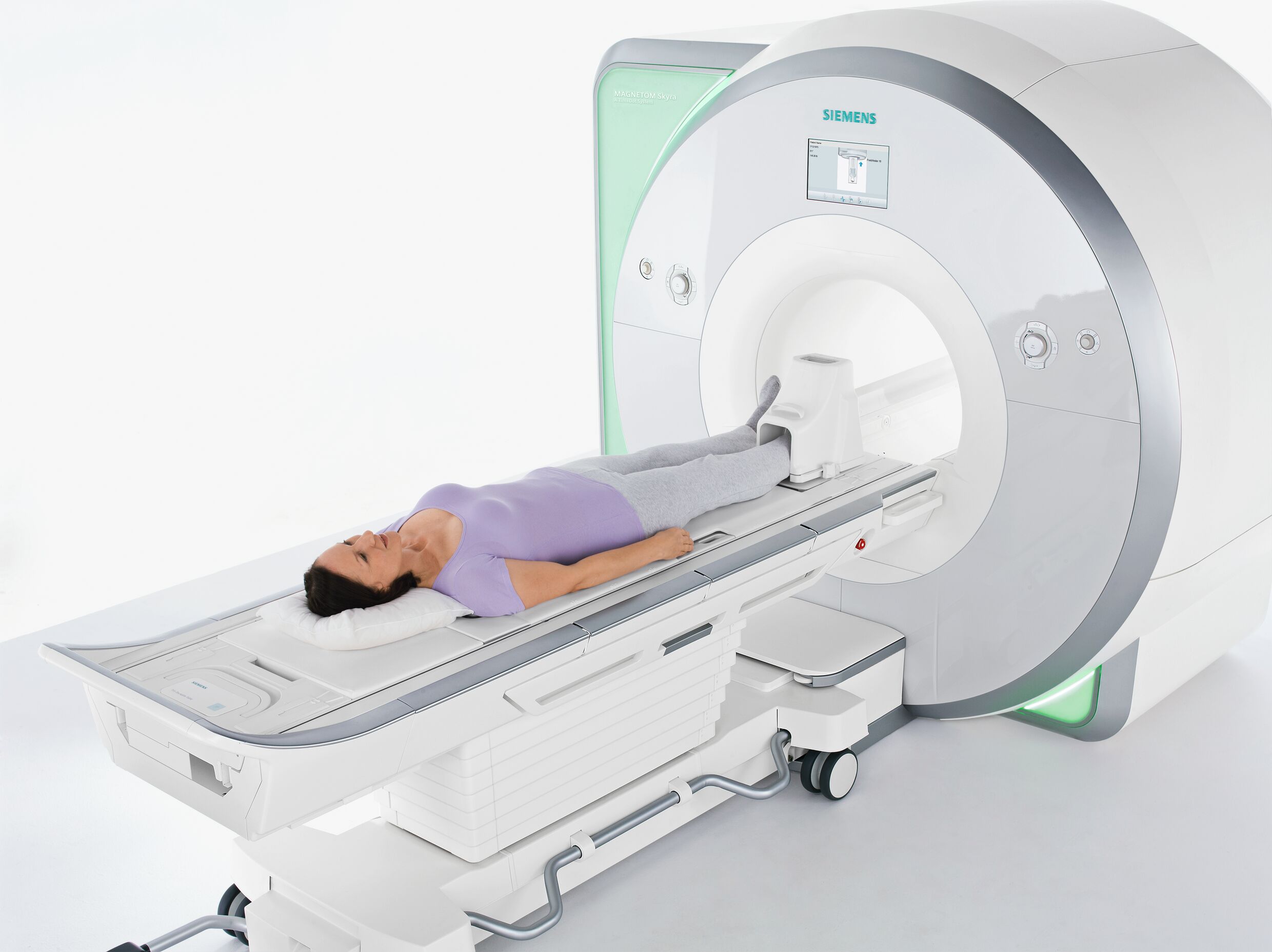
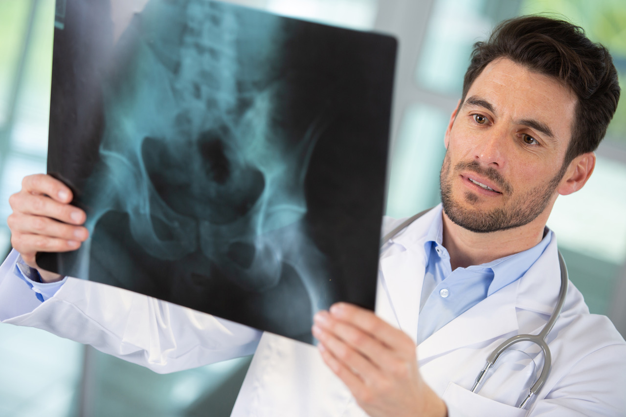
Osteoarticular MRI

Osteoarticular MRI is a non-invasive imaging test that uses a powerful magnetic field to obtain detailed images of bones, joints, ligaments, tendons and muscles. This examination is essential for diagnosing and evaluating joint and musculoskeletal pathologies, such as osteoarthritis, ligament tears or inflammation.
Breast MRI
Advanced imaging using a powerful magnetic field and radio to obtain highly detailed images of breast tissue. This examination is particularly useful for detecting abnormalities not visible on mammography or ultrasound and for assessing the extent of breast cancer.
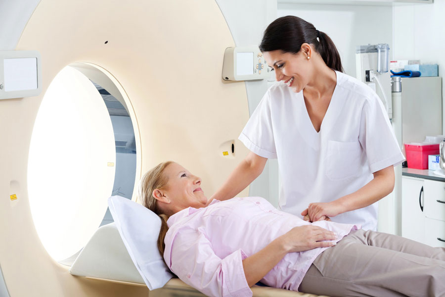
Brain and pre-brain MRI

Brain and pre-brain MRI is an imaging test that uses strong magnetic fields to obtain detailed images of the brain and surrounding structures. This examination is crucial for diagnosing neurological pathologies, evaluating strokes, brain tumors or abnormalities of cerebral vessels.
Cardiac MRI
Cardiac MRI is an advanced imaging examination that uses a powerful magnetic field and radio waves to obtain detailed images of the heart and surrounding structures. This examination allows for a precise evaluation of the heart muscle, valves, and coronary vessels, and is essential for diagnosing complex heart conditions, assessing the after-effects of a heart attack, or planning surgical interventions.
Guided infiltrations
Guided injections are therapeutic procedures that use imaging (MRI, CT scan, or ultrasound) to precisely guide the injection of anti-inflammatory drugs or anesthetics into a joint or near a nerve. These injections are performed to relieve pain, reduce inflammation, or improve joint mobility.
Urogenital ultrasound

Urogenital ultrasound is a non-invasive examination that uses sound waves to visualize the male genital and urinary organs, such as the bladder, prostate, testicles, and kidneys. This examination is often used to evaluate urological symptoms, detect abnormalities, or monitor pre-existing conditions
Breast ultrasound
Breast ultrasound: Breast ultrasound uses sound waves to produce real-time images of breast tissue. This examination is particularly useful for examining masses detected during a mammogram or for visualizing tissue that is difficult to see on X-ray images
Lung Cancer Screening

Lung cancer screening Lung cancer screening relies on the use of low-dose chest scans to detect early signs of lung abnormalities, often asymptomatic, in people at risk.
vascular CT gated

Gated vascular CT uses X-ray scanners and cardiac cycle synchronization to obtain precise images of blood vessels. This examination allows for a detailed evaluation of arteries and veins, minimizing artifacts due to cardiac motion, and is essential for diagnosing vascular diseases and planning interventions
Cardiac and coronary multiphase CT

Multiphase cardiac and coronary CT is an imaging technique that uses X-ray scanners to obtain multiphase images of the heart and coronary vessels. This method allows for detailed visualization of the coronary arteries and cardiac structures and can help evaluate coronary artery disease or heart abnormalities.
High-resolution coronary angiography

High-resolution coronary angiography (HRCA) is a precise imaging examination that uses X-ray scanners to obtain detailed images of the coronary arteries. This technique allows for meticulous visualization of the coronary arteries and is used to diagnose coronary artery disease, plan interventions, and assess the condition of stents or coronary artery bypass grafts.
Prostate biopsy using UroNav

UroNav Prostate Biopsy: A UroNav prostate biopsy is an advanced procedure used to diagnose prostate cancer. UroNav combines detailed prostate MRI images with a real-time guidance system to precisely locate suspicious areas and collect tissue samples.
